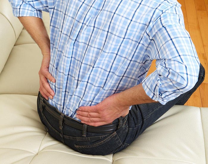
Renal Colic
Renal colic is pain caused by a urinary tract stone (urolithiasis).
Stones can vary significantly in size. Most stones occur due to a buildup of minerals or other substances, such as uric acid, which stick together in the urine, creating a hard mass.
There are many treatment options for urinary stones. Many stones pass without surgical intervention, making managing renal colic a primary concern during treatment.
Symptoms

Renal colic pain may be experiences at the sides of the body between the lower ribs and hips.
Some small stones cause only mild renal colic, and a person may pass them in the urine without much discomfort.
Larger stones can cause excruciating pain, especially if they get stuck and block any small points in the urinary tract.
The most common presentation of renal colic is pain that occurs on the affected side of the body between the lower ribs and hip that radiates to the lower abdomen and groin.

The pain tends to come in waves that can last from 20 to 60 minutes before subsiding until the next wave.
Other symptoms that typically occur alongside renal colic include:
- pain or difficulty urinating
- blood in the urine which may give it a pinkish, red, or brown color
- foul-smelling urine
- nausea
- vomiting
- small particles in the urine
- feeling a constant urgent need to urinate
- cloudy urine
Anyone experiencing the following symptoms in addition to renal colic should contact emergency medical services immediately:
- complete inability to urinate
- uncontrollable vomiting
- a fever over 101°F
Causes
Urinary stones can be made up of numerous chemicals and minerals caused by a few different risk factors. Risk factors for developing a urinary stone include:
- extra calcium in the urine
- diseases of the gastrointestinal (GI) tract, such as Crohn’s disease or ulcerative colitis
- gout, which is caused by an excess of uric acid
- certain medications
- cystinuria
- obesity
- surgeries in the GI tract, such as gastric bypass surgery
- dehydration
- a family history of urolithiasis
Treatment and types

A close up image of a calcium oxalate stone.
Doctors will often use blood tests to check for increased levels of stone-forming substances in a person’s body. An imaging test such as a plain film X-ray, a computed tomography (CT) scan, or ultrasound can help locate any significant stones in the urinary tract.
In fact, up to 80 percent of stones will pass out of the body in the urine. Doctors will recommend proper hydration and may prescribe pain-relieving medications to help deal with the pain while monitoring the stone until it passes.
There is a range of procedures to help remove larger stones and relieve renal colic.
- Ureteroscopy guided laser stone fragmentation: This is an invasive surgical procedure where a doctor inserts a thin scope with a light and camera on it into the urinary tract to locate the stone and remove it.
- Extracorporeal shock wave lithotripsy (ESWL): A noninvasive treatment, ESWL is the process of aiming small soundwaves at the kidneys to break up stones into tiny pieces. These fragments are then passed in the urine.

- Percutaneous nephrolithotomy: Percutaneous nephrolithotomy is typically done under general anesthesia. It is the process of entering the kidney through a small cut in the back and using a lighted scope and small instruments to remove the stone.
- Stent placement: Sometimes, doctors will place a thin tube into a person’s ureter to help relieve the obstruction and promote the passing of stones.

Prevention

Alongside increasing the fluid intake, doctors may recommend citrus fruits in the diet to help prevent renal colic.

It is possible for stones in the urinary tract to happen again after successful treatment. Taking preventative measures can help avoid developing stones in the future and reduce symptoms of renal colic.
